Above Right:
(L to R:)
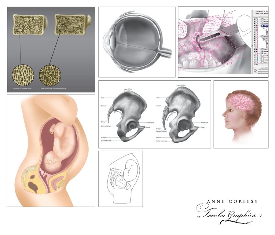
1. Orthopaedic illustration: cross section of vertebra; normal versus bone affected by osteoporosis. Digital illustration. Adobe Illustrator & Photoshop.
2. Ophthalmic illustration: eye cross-section. Digital illustration. Adobe Illustrator & Photoshop.
3. Surgical sequence (one image only here): maxillofacial surgery, Le Fort 1 osteotomy procedure. Digital illustration. Adobe Illustrator Gradient Mesh.
4. Obstetric illustration: cross section showing fetus in utero. Digital illustration. Adobe Illustrator & Photoshop.
5. Orthopaedic illustration showing the anatomical differences between the male and female pelvis. Commission for Wellcome Collection, London, UK. Digital illustration. Adobe Illustrator & Photoshop.
6. Anatomical illustration showing part of the brain. Digital illustration. Adobe Illustrator & Photoshop.
7. Obstetric illustration to show the internal and external conjugates and engagement of the fetal head. Digital illustration. Adobe Illustrator.
(L to R:)
1. Orthopaedic illustration: cross section of vertebra; normal versus bone affected by osteoporosis. Digital illustration. Adobe Illustrator & Photoshop.
2. Ophthalmic illustration: eye cross-section. Digital illustration. Adobe Illustrator & Photoshop.
3. Surgical sequence (one image only here): maxillofacial surgery, Le Fort 1 osteotomy procedure. Digital illustration. Adobe Illustrator Gradient Mesh.
4. Obstetric illustration: cross section showing fetus in utero. Digital illustration. Adobe Illustrator & Photoshop.
5. Orthopaedic illustration showing the anatomical differences between the male and female pelvis. Commission for Wellcome Collection, London, UK. Digital illustration. Adobe Illustrator & Photoshop.
6. Anatomical illustration showing part of the brain. Digital illustration. Adobe Illustrator & Photoshop.
7. Obstetric illustration to show the internal and external conjugates and engagement of the fetal head. Digital illustration. Adobe Illustrator.